


Optical Coherence Tomography RS-1 Glauvas
Optical Coherence Tomography
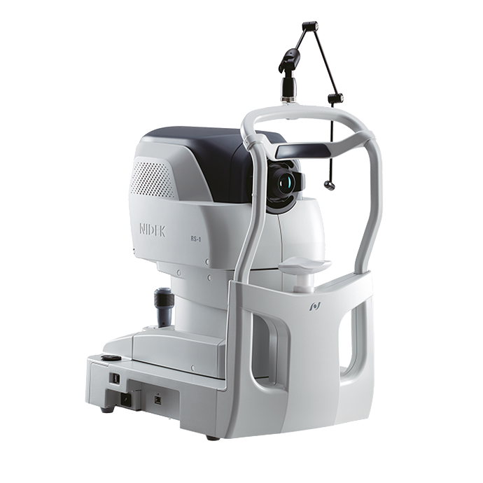
The device delivers ultra high-speed imaging with up to 250,000 A-scans per second, providing wide, deep, and high-resolution visualization. This capability enables highly detailed analysis for advanced ophthalmic diagnostics. Its effortless operation and intuitive interpretation streamline clinical workflow. Powerful analytical tools support in-depth evaluation and comparison of images for enhanced clinical insights.
Optical Coherence Tomography RS-1 Glauvas
NIDEK
The incorporation of 250,000 A-scans per second significantly reduces image capture time and accelerates the diagnostic workflow. High-speed imaging also minimizes patient fixation errors, leading to greater image clarity and improved comfort. With RS-1 Glauvas, a single B-scan clearly visualizes the area from the optic nerve head to the temporal vascular arcade. The 4.2 mm depth B-scan enables precise imaging of the oblate retinal shape commonly seen in myopic eyes. Improvements in AngioScan OCT-Angiography provide wider and sharper images for detailed assessment of the chorioretinal microvasculature. Image acquisition is simplified by automated functions. For glaucoma analysis in myopic patients, deep learning segmentation (DL segmentation) reduces the likelihood of false positives. The new Structural Normality Map (SN Map) enhances diagnostic confidence in early-stage disease by clearly displaying deviations from normal tissue structure.


Detailed information
250,000 A-scans/s high-speed imaging
The incorporation of 250,000 A-scans/s accelerates your workflow by reducing capture time. The high-speed imaging also addresses patient fixation errors thus contributing to greater image clarity and patient comfort.
Wide, deep, high-resolution imaging
With RS-1 Glauvas, a single B-scan image clearly presents the area from the optic nerve head to the temporal vascular arcade, and the 4.2 mm depth B-scan imaging readily captures the oblate retinal shape of myopic eyes. Improvements in AngioScan OCT-Angiography include wider and clearer images for assessing chorioretinal microvasculature.
Effortless operation and interpretation
Easy image capture with automated functions
The auto alignment and auto switch functions allow anyone to effortlessly capture images. Operators need to only adjust the chinrest height and click Optimize and Capture.
Advanced analytics
Glaucoma analysis in myopia
The long axial length normative database*1 presents analysis with axial length compensation, allowing for a more accurate glaucoma assessment in patients with axial myopia. The OCT Viewer automatically switches to this database as required, by using the axial length*2 which is a parameter for scan width correction.
Less false positives with deep learning segmentation (DL segmentation)
The accuracy of segmentation affects the outcomes of glaucoma analysis. DL segmentation reduces artifacts and errors in the normative database and thickness maps even in eyes with opacities, thus decreasing false positives and enhancing clinic efficiency by reducing unnecessary follow-up visits. Additionally, the scan width correction allows precise analytics based on the patient’s axial length*2.
*1 Data was collected from a sample of Asian patients.
*2 The value of the axial length is obtained based on the results of the OCT image capture and differs from the actual measured value of the axial length.
Structural Normality Map (SN Map) improving diagnostic confidence on early detection
The DL segmentation provides an SN Map that presents structural abnormalities and changes. This functionality aids clinicians in detecting minute structural changes at a glance, contributing to greater diagnostic confidence even for early signs of retinal changes.
