


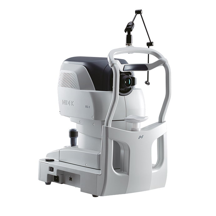
Optical Coherence Tomography RS-1 Glauvas
Օպտիկական Կոհերենտ Տոմոգրաֆիա
Սարքը ապահովում է բարձր արագությամբ պատկերում՝ մինչև 250,000 A-սքան վայրկյանում, որը թույլ է տալիս ստանալ լայնածավալ, խորքային և բարձր լուծաչափով պատկերներ։ Այս հնարավորությունն էականորեն բարձրացնում է ախտորոշման ճշգրտությունը՝ հատկապես մանրամասն կառուցվածքների գնահատման դեպքում։ Կառավարման գործընթացը դյուրին է, իսկ արդյունքների մեկնաբանությունը՝ հստակ և հասկանալի, ինչը հեշտացնում է ինչպես օպերատորի, այնպես էլ բժշկի աշխատանքը։ Ընդլայնված վերլուծական գործիքակազմը թույլ է տալիս կատարել խորքային գնահատում, համեմատական վերլուծություն և հետևել դինամիկ փոփոխություններին ժամանակի ընթացքում։
Optical Coherence Tomography RS-1 Glauvas
NIDEK
Պատկերահանման արագության բարձրացումը՝ մինչև 250,000 A-սքան վայրկյանում, զգալիորեն կրճատում է սքանավորման ժամանակը և արագացնում է հետազոտման գործընթացը։ Բարձր արագությամբ պատկերման շնորհիվ նվազում են պացիենտի ֆիքսացիոն սխալները, ինչը ապահովում է ավելի հստակ պատկերներ և բարձրացնում է ընդհանուր հարմարավետությունը։ RS-1 Glauvas սարքը թույլ է տալիս մեկ B-սքանով հստակ պատկերել տարածքը՝ սկսած օպտիկ նյարդի գլխից մինչև ժամանակային անոթային աղեղ։ 4.2 մմ խորությամբ B-սքանը հարմար է նաև միոպիկ աչքի ռետինայի օվալաձև կառուցվածքը լիարժեք նկարագրելու համար։ AngioScan OCT-Angiography համակարգում կատարված բարելավումները թույլ են տալիս ստանալ ավելի լայն և բարձր հստակությամբ պատկերներ՝ խոռոչաթաղանթային մանրանոթային ցանցի գնահատման համար։ Պատկերի հեշտ և արագ ստացումը հնարավոր է ավտոմատացված ֆունկցիաների շնորհիվ։ Միոպիկ գլաուկոմայի վերլուծության համար կիրառվում է խորքային ուսուցման սեգմենտացիա (DL segmentation)՝ նվազեցնելով կեղծ դրական արդյունքների հավանականությունը։ Նոր կառուցվածքային նորմալության քարտեզը (SN Map) բարձրացնում է վաղ ախտորոշման վստահությունը՝ ներկայացնելով կառուցվածքային շեղումները նորմայից։


Մանրամասն տեղեկատվություն
250,000 A-scans/s high-speed imaging
The incorporation of 250,000 A-scans/s accelerates your workflow by reducing capture time. The high-speed imaging also addresses patient fixation errors thus contributing to greater image clarity and patient comfort.
Wide, deep, high-resolution imaging
With RS-1 Glauvas, a single B-scan image clearly presents the area from the optic nerve head to the temporal vascular arcade, and the 4.2 mm depth B-scan imaging readily captures the oblate retinal shape of myopic eyes. Improvements in AngioScan OCT-Angiography include wider and clearer images for assessing chorioretinal microvasculature.
Effortless operation and interpretation
Easy image capture with automated functions
The auto alignment and auto switch functions allow anyone to effortlessly capture images. Operators need to only adjust the chinrest height and click Optimize and Capture.
Advanced analytics
Glaucoma analysis in myopia
The long axial length normative database*1 presents analysis with axial length compensation, allowing for a more accurate glaucoma assessment in patients with axial myopia. The OCT Viewer automatically switches to this database as required, by using the axial length*2 which is a parameter for scan width correction.
Less false positives with deep learning segmentation (DL segmentation)
The accuracy of segmentation affects the outcomes of glaucoma analysis. DL segmentation reduces artifacts and errors in the normative database and thickness maps even in eyes with opacities, thus decreasing false positives and enhancing clinic efficiency by reducing unnecessary follow-up visits. Additionally, the scan width correction allows precise analytics based on the patient’s axial length*2.
*1 Data was collected from a sample of Asian patients.
*2 The value of the axial length is obtained based on the results of the OCT image capture and differs from the actual measured value of the axial length.
Structural Normality Map (SN Map) improving diagnostic confidence on early detection
The DL segmentation provides an SN Map that presents structural abnormalities and changes. This functionality aids clinicians in detecting minute structural changes at a glance, contributing to greater diagnostic confidence even for early signs of retinal changes.
